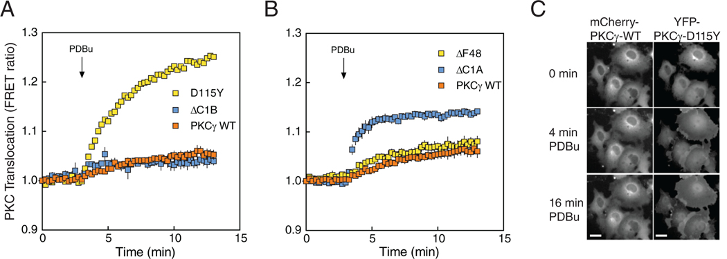
Figure 3. SCA14 mutations affect translocation of PKCγ.
(A) COS7 cells were co-transfected with MyrPalm-CFP and YFP-tagged WT PKCγ (orange), PKCγ D115Y (yellow), or PKCγ ΔC1B (blue). Rate of translocation to plasma membrane was monitored by measuring FRET/CFP ratio changes after addition of 200 nM PDBu. Data were normalized to the starting point (1.0) and are representative of two independent experiments, N ≥ 22 cells per condition. (B) COS7 cells were co-transfected with MyrPalm-CFP and YFP-tagged ΔF48 or ΔC1A. Data are mean ± S.E.M. from at least three independent experiments, N ≥ 23 cells per condition. (C) COS7 cells were co-transfected with mCherry-tagged WT PKCγ and YFP-tagged PKCγ D115Y. Localization of mCherry-PKCγ (WT; left) and YFP-PKCγ-D115Y (right) in the same cells under basal conditions and after addition of 200 nM PDBu was observed by fluorescence microscopy. Images are representative of three independent experiments. Scale bar = 20 μm.

