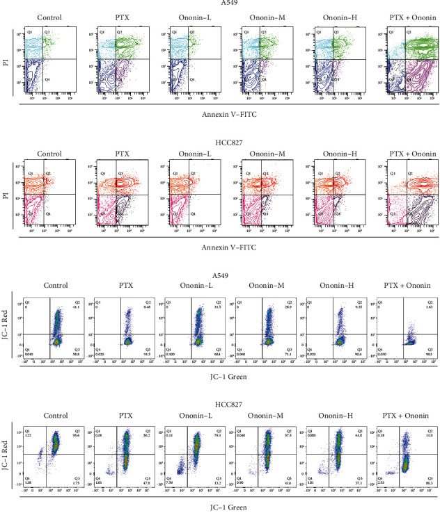
Figure 2.

Ononin stimulates NSCLC apoptotic ability. (a, b) The viable cell population was displayed in the bottom left quadrant (Q3), the early apoptotic cells in the bottom right quadrant (Q4), and the late apoptotic cells in the top right quadrant (Q4) of the dual parametric dot plots integrating Annexin V-FITC and PI fluorescence (Q2). (c, d) The MMP level was detected by a flow cytometry with fluorescent dye JC-1. Cytogram plots of control and samples incubated with PTX (1 μM, positive control) or ononin (0.3, 1, and 3 μM) or the combination of PTX (1 μM) and ononin (1 μM) for 48 h, respectively. The horizontal axis showed the green fluorescence (JC-1 monomer), and the vertical axis indicated red fluorescence (JC-1 aggregates).
