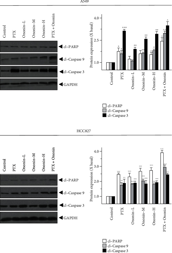
Figure 4.

Ononin initiates apoptosis biomarker expressions. Cultured A549 or HCC827 cells were incubated with PTX (1 μM) or ononin (0.3, 1, and 3 μM) or the combination of PTX (1 μM) and ononin (1 μM) for 2 days, and the target proteins were detected by western blot. The quantification of the target protein was calculated by a densitometer. The values are expressed as the fold of changes (X basal), in mean ± SEM, where n = 6. ∗p < 0.05, ∗∗p < 0.01, and ∗∗∗p < 0.001 when compared to the control group. When compared to the PTX group, significant values were indicated by ^p < 0.05, ^^p < 0.01, and ^^^p < 0.001.
