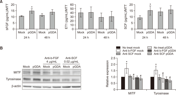
Fig. 1.
Expression levels of bFGF and SCF are increased in keratinocytes with GDA overexpression. (A) Concentrations of bFGF, SCF, and ET-1 in primary cultured human keratinocytes with or without GDA overexpression based on ELISA. (B) Western blot analysis of relative ratios of protein levels of MITF and tyrosinase in melanocytes treated with culture supernatants from GDA-overexpressing keratinocytes in the presence or absence of anti-bFGF and anti-SCF antibodies. β-Actin was used as an internal control for Western blot analysis. Data are as present means ± SD of four independent experiments. *p<0.05 vs. control keratinocytes, #p<0.05 vs. non-treated GDA-overexpressing keratinocytes.

