Fig. 1.

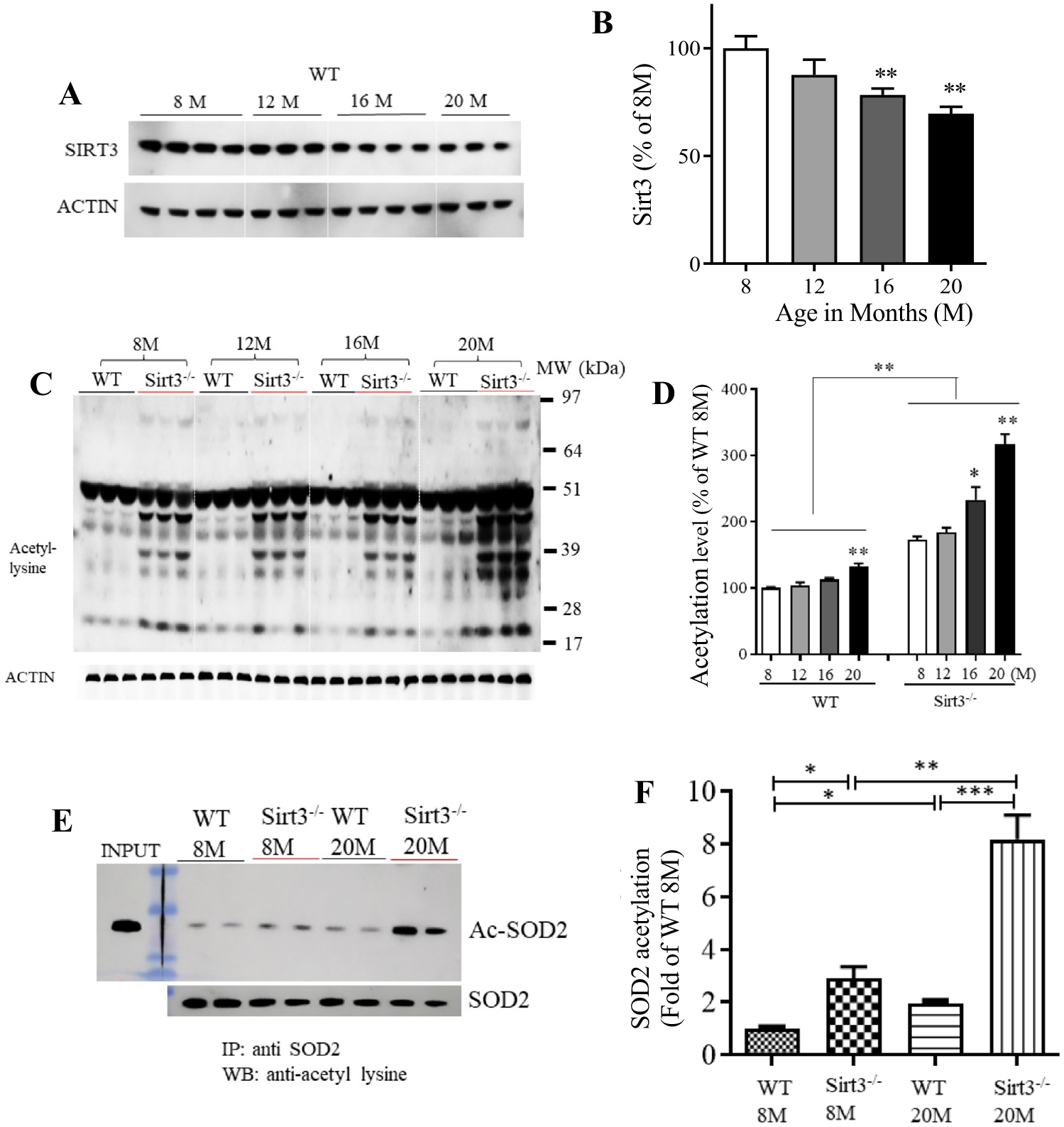
SIRT3 levels decrease and mitochondrial protein acetylation increases in the cerebral cortex during aging. A Immunoblot analysis of SIRT3 protein in cortical tissue from wild type (WT; Sirt3+/+) mice at different ages (8, 12, 16 and 20 month). Actin was used as a loading control. B SIRT3 levels were normalized to actin protein levels and expressed as a percentage of the value for 8-month-old mice. Values are mean ± SEM (n = 7 mice). **p < 0.01 (One-way ANOVA with Tukey post hoc test). C Immunoblot analysis of acetylated proteins detected using an acetyl-l-lysine antibody in protein samples prepared from cortical tissues of Sirt3+/+ and Sirt3−/− mice of 8, 12, 16 and 20 months (M) of age. Actin was used as a loading control. D Total protein acetylation levels (densitometric analysis of immunoreactivity in the entire lane) were normalized to actin protein levels and expressed as a percentage of the value for Sirt3+/+ 8-month-old mice. Values are mean ± SEM (n = 6 mice). *p < 0.05; **p < 0.01 (one-way ANOVA with Tukey post hoc test). E Proteins in lysates of cortical tissues from young (8 months) and old (20 months) Sirt3+/+ and Sirt3−/− mice were immunoprecipitated with an SOD2 or antibody and subjected to immunoblot analysis with antibodies to acetyl-lysine (Ac-SOD2). The blots were re-probed with SOD2 antibody to control for total SOD2 level. Input refers to the direct immunoblotting of tissue lysate using an antibody against SOD2. F Acetylated SOD2 level normalized to the SOD2 level and expressed as percentage of the value for 8-month-old Sirt3+/+ mice. Values are mean ± SEM (n = 6 mice). *p < 0.05; **p < 0.01 (one-way ANOVA with Tukey post hoc test)
