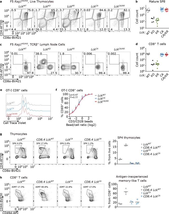
Extended Data Fig. 8. Characterization of CD8+ T cells in the Lck variant mice.
(a-d) Thymi (a-b) and LNs (c-d) of indicated Lck variant F5 Rag1KO/KO mice were analyzed by flow cytometry. (a) Expression of CD4 and CD8α in representative mice (gated on viable cells). (b) Counts of mature SP8 (viable CD4− CD8α+ CD24− TCRβ+) thymocytes. Individual mice and medians are shown. n = 10 LckWT/WT in 7 independent experiments, 5 LckKO/KO in 3 independent experiments, 9 LckKR/KR, 13 LckCA/CA and LckCA/KR in 8 independent experiments. (c) Expression of CD4 and CD8α in representative mice (gated on viable cells). (d) Total numbers of CD8+ T cells in LNs. Individual mice and medians are shown. n = 10 LckWT/WT in 7 independent experiments, 5 LckKO/KO in 3 independent experiments, 9 LckKR/KR, 12 LckCA/CA and 13 LckCA/KR in 8 independent experiments. (e-f) LN cells from indicated Lck variant OT-I mice were loaded with Cell Trace violet (CTV) and stimulated with anti-CD3/CD28 beads. (e) The proliferation was evaluated based on the CTV dilution at 72 hours after activation by flow cytometry. A representative experiment/mice out of 4 in total. (f) Upregulation of CD69 was analyzed by flow cytometry at 16 hours after activation. Mean + s.e.m. n = 3 independent experiments/mice. Differences in the EC50 and/or maximum of the fitted non-linear regression curves were tested using extra sum-of-squares F test. F and p values are shown. (g) Thymocytes from indicated Lck variant CD8 WT or CD8.4 OT-I mice were analyzed by flow cytometry. Representative mice and the frequencies of SP4 T cells from 3 independent experiments/mice are shown. (h) LN cells from indicated Lck variant CD8 WT or CD8.4 OT-I mice were analyzed by flow cytometry. Representative mice and the frequencies of CD44+ CD49d− antigen-inexperienced memory-like cells (individual values and means) from 3 independent experiments/mice are shown.

