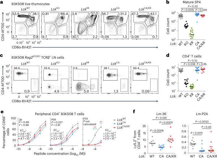
Fig. 6. Kinase-dependent and kinase-independent roles of CD4-bound LCK in T cell responses.
a–d, Thymi (a and b) and LNs (c and d) of indicated B3K508 mice were analyzed by flow cytometry. Statistical significance was calculated using a Mann–Whitney test. a, Expression of CD4 and CD8 in representative mice (gated on viable cells). b, Numbers of mSP4 (viable CD4−CD8α+CD24−TCRβ+) T cells; LckWT/WT: n = 24 mice in 10 independent experiments; LckKO/KO: n = 12 mice in 7 independent experiments; LckKR/KR: n = 13 mice in 6 independent experiments; LckCA/CA: n = 19 mice in 8 independent experiments; LckCA/KR: n = 10 mice in 3 independent experiments. Individual mice and medians are shown. c, CD4+ and CD8+ T cells in representative mice (gated on viable TCRβ+ cells). d, Numbers of CD4+ T cells in individual mice and medians are shown; LckWT/WT: n = 26 mice in 11 independent experiments; LckKO/KO: n = 11 mice in 7 independent experiments; LckKR/KR: n = 13 mice in 6 independent experiments; LckCA/CA: n = 20 mice in 9 independent experiments; LckCA/KR: n = 10 mice in 3 independent experiments. e, T cells isolated from LNs of indicated Lck-variant B3K508 mice were activated ex vivo with splenocytes from Ly5.1 mice loaded with 3K peptide or APLs with decreasing affinity (3K > P5R > P2A) overnight and analyzed for the expression of CD69 by flow cytometry. Mean values + s.e.m. are shown; number of independent experiments/mice: n = 7 (LckCA/KR) or 8 (other strains) for 3K, 5 for P5R and 6 (LckCA/KR) or 8 (other strains) for P2A. Differences in the EC50 and/or maximum of the fitted non-linear regression curves were tested using an extra sum of squares F-test. F, P and EC50 values are shown. The significance of the differences at individual concentrations was calculated using a Mann–Whitney test and is displayed between LckWT/WT and LckCA/CA mice (blue stars) and LckWT/WT and LckCA/KR mice (red stars); *P < 0.05; **P < 0.01; no symbol, P > 0.05 (Supplementary Table 5). f, Indicated Lck-variant B3K508 Rag2KO/KO T cells were transferred into Ly5.1 mice followed by infection with Lm expressing 3K or P2A. Expansion of B3K508 T cells was measured as percentage among total CD4+ T cells on day 5 after infection by flow cytometry. Results for individual mice and medians are shown; n = 8 LckWT/WT, n = 11 LckCA/CA and n = 10 LckCA/KR mice from three independent experiments for Lm-3K; n = 12 LckWT/WT, n = 13 LckCA/CA and n = 13 LckCA/KR mice from three independent experiments for Lm-P2A. Statistical significance was calculated using a Mann–Whitney test.

