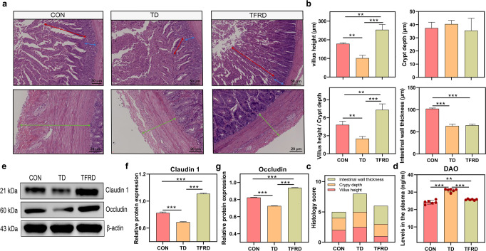
Fig. 5. Duodenal morphology and mucosal barrier function after TFRD supplementation.
a Representative images of HE-stained sections of the duodenum. Red arrow, villus height. Blue arrow, crypt depth. Cyan arrow, intestinal wall thickness (scale bars: top, 50 µm; bottom, 20 µm). b Statistical analyses of the villus height, crypt depth, the ratio of villus height to crypt depth and intestinal wall thickness. c Stacked bar graph showing the differences in the total histology scores and individual histological criteria scores between the three groups. d Plasma DAO levels (n = 6 broilers per group). e–g Western blots showing Claudin 1 and Occludin expression in the duodenum. *p < 0.05, **p < 0.01, and ***p < 0.001. One-way ANOVA with LSD post hoc test. The data are presented as the means ± SD.

