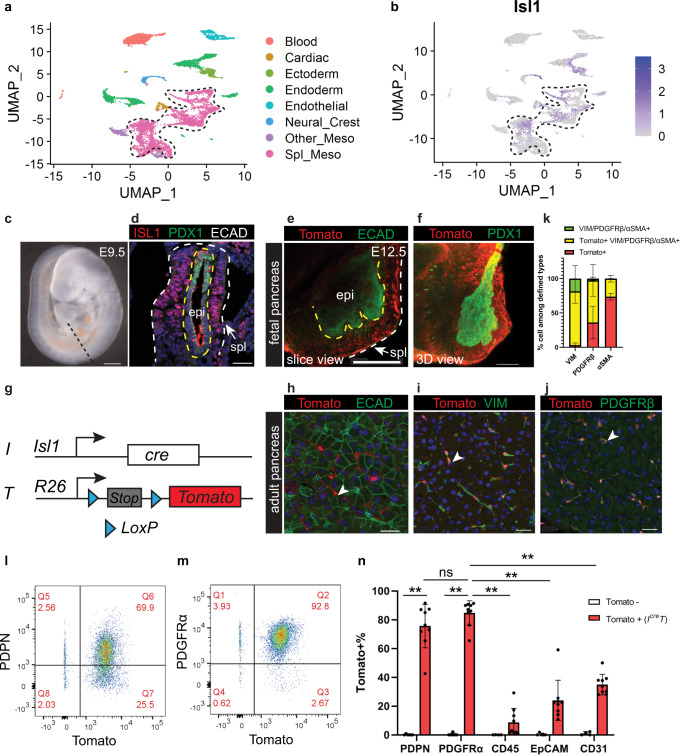
Fig. 2. Tissue-resident fibroblasts in the adult pancreas are derived from ISL1 expressing splanchnic mesenchyme during fetal development.
a Clustering analysis of single-cell transcriptome profiles of E9.5 mouse foregut cells. b Isl1 expression projected on clusters in (a). Dashed lines delineate the splanchnic mesenchyme cluster. c Lateral view of an E9.5 mouse embryo. Dashed line indicates a transverse section at the pancreatic level. d Immunostaining on mouse foregut indicated in (c). n = 3 embryos. e, f Whole mount immunostaining of a dissected pancreas. Yellow dashed lines delineate the epithelium, while the yellow and the white lines delineate the splanchnic mesenchyme. n = 3 pancreata. g Schematic of the IcreT mouse model. h–k Co-immunostaining of the adult pancreas and quantification of cells that are either single positive or double positive for defined markers. n = 5 mice. l–n Flow cytometry analysis of IcreT adult pancreas and quantification of Tomato+ cells within populations defined by markers. Tomato-, n = 5 mice; IcreT, n = 9 mice. Data are mean ± SD. Two-way ANOVA test with Tukey’s multiple comparisons was performed. ** indicates p < 0.0001. ns, not significant, p = 0.547. E, embryonic day; epi, epithelium; meso, mesoderm; spl, splanchnic mesenchyme. Arrowheads indicate Tomato+ cells. Scale bars in (c–f): 100 μm; in (h–j): 30 μm. Source data are provided as a Source data file.

