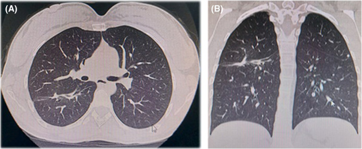
FIGURE 3.

(A) Axial computed tomography view, showing thickening of the major fissure of the right lung associated with a small hyperdense image of irregular subpleural shape adjacent to the major fissure of the lower lobe, as residual changes in relation to pathological and surgical history. (B) Coronal computed tomography view.
