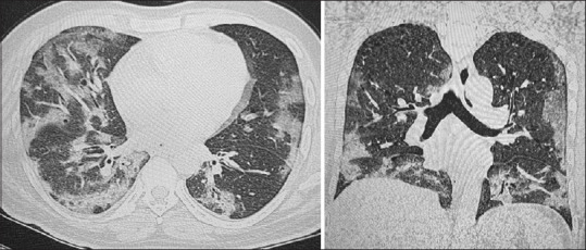
Figure 3.

Axial and coronal images from the HRCT Chest of a 44-year-old male patient. There is moderate lung involvement with multiple, peripheral patchy areas of ground glass attenuation and septal thickening. The findings were more prominent in the lower lobes bilaterally. The CT severity score was 18/25
