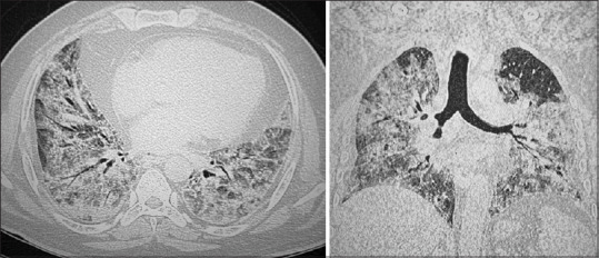
Figure 4.

Axial and coronal images of HRCT chest of a 61–year-old male patient showing extensive, confluent areas of ground glass attenuation and consolidations diffusely involving both lungs interspersed with septal thickening. Reported as ARDS and the CT severity score was 24/25
