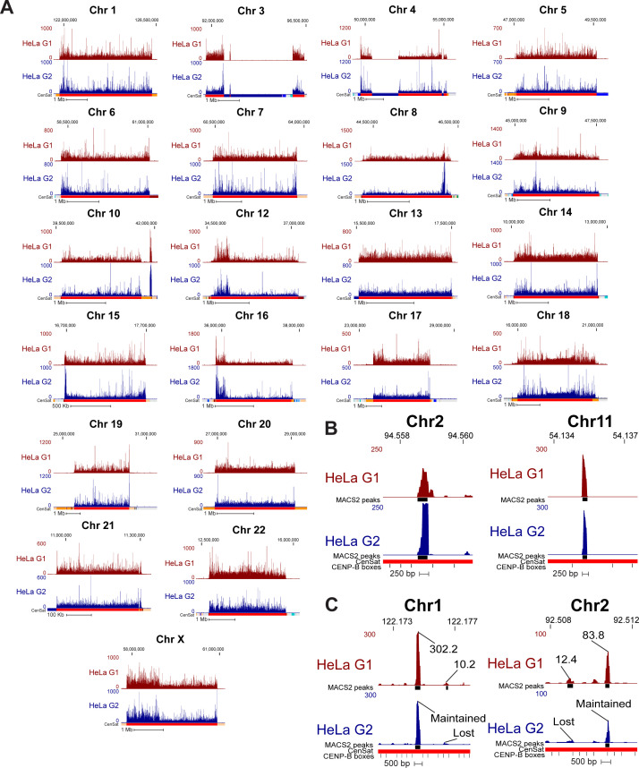
Figure S5. CENP-A position is maintained through DNA replication on the T2T assembly.
(A) Mapping of CENP-A-bound reads in HeLa G1 (top, maroon) and G2 (bottom, dark blue) cells to the T2T assembly at all centromeres (excluding centromeres 2 and 11, presented in Fig 4). (B) High-resolution views of CENP-A peaks depicting the position of single CENP-A nucleosomes that are maintained from G1 to G2. (C) High-resolution views of CENP-A peaks that are not maintained from G1 to G2 next to G1 peaks that are maintained into G2. Lost G1 peaks are enriched for CENP-A at a low level. Peak values are enrichment values for CENP-A over input reported by MACS2 (P < 0.00001, ≥10-fold enrichment).

