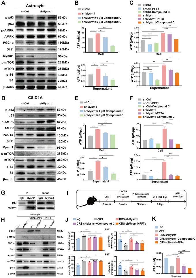
Figure 6.

Mysm1 knockdown activates the p53/AMPK pathway and enhances ATP generation in astrocytes. A) Western blot of indicative proteins in primary astrocytes transduced with control (shCtrl) and Mysm1 knockdown (shMysm1) virus. B,C) ATP levels in astrocytes (top) and the supernatant (bottom) with the indicative treatment. D) Western blot of indicative proteins in C8‐D1A cells transduced with control (shCtrl) and Mysm1 knockdown (shMysm1) virus. E,F) ATP levels in C8‐D1A cells (top) and the supernatant (bottom) with the indicative treatment. G) The interaction between p53 and Mysm1 determined by Co‐immunoprecipitation (Co‐IP) in astrocytes. H) Western blot results in control and Mysm1 knockdown C8‐D1A cells stimulated with Compound C or PFTα. I) Schematic diagram of Compound C or PFTα treatment in Mysm1 knockdown CRS mice and the behavior tests. J) The TST and FST of mice with the indicative treatments in the MHb. K) ATP content in the serum of Compound C‐ and PFTα‐treated Mysm1 knockdown CRS mice and the control mice (NC). Data represent the mean ± SEM; ns, not significant; *p < 0.05, **p < 0.01, ***p < 0.001, and ****p < 0.0001.
