Fig. 1.
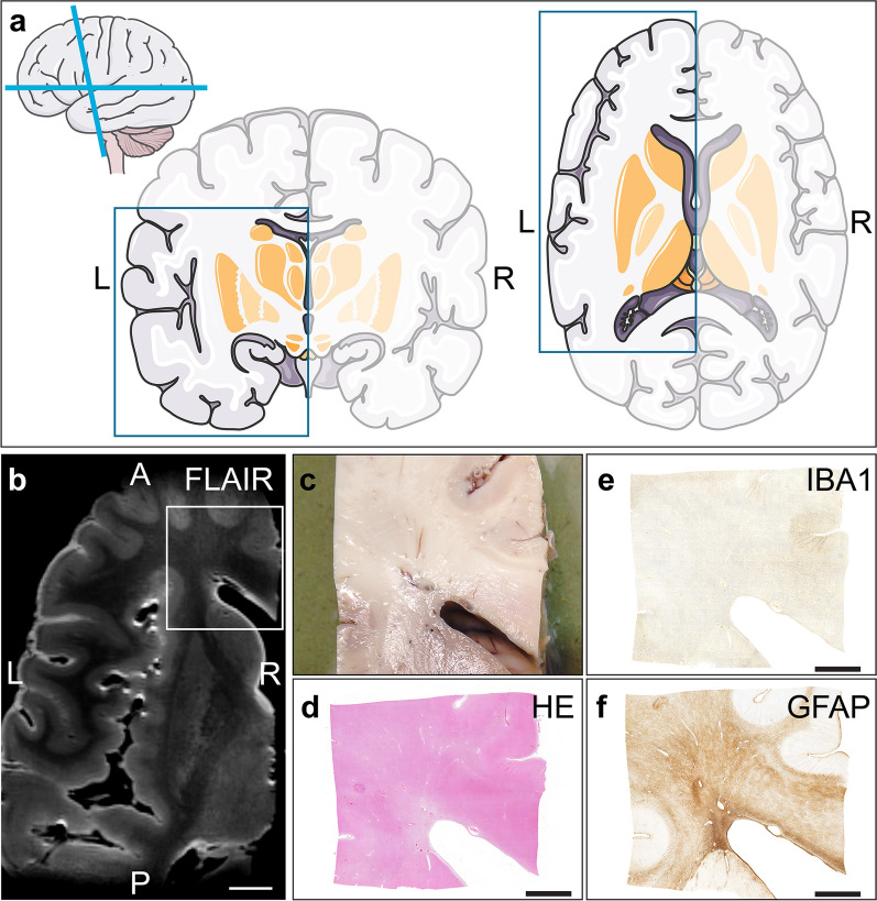
Tissue preparation. The left hemisphere was divided into dorsal and ventral part for HF (7 Tesla) MRI scanning. a Schematic brain images. The blue squares illustrate the dimensions of the ventral part of the left hemisphere. b Corresponding HF MRI fluid-attenuated inversion recovery (FLAIR) axial slab (white scale bar = 1 cm). The white rectangle illustrates the tissue blocks of approximately 2 × 2 × 0.5 cm taken from the (c) biopsy. (Immuno-)histopathology of (d) haematoxylin/eosin (HE), (e) ionized calcium-binding adapter molecule 1 (IBA1) to detect macrophages and microglia, and (f) glial fibrillary acidic protein (GFAP) to detect astrocytes. This figure was partly generated using “Neurology” images from Servier Medical Art (https://smart.servier.com), licensed under a Creative Commons Attribution 3.0 Unported License. (black scale bar = 0.5 cm) (L left, R Right, A Anterior, P Posterior)

