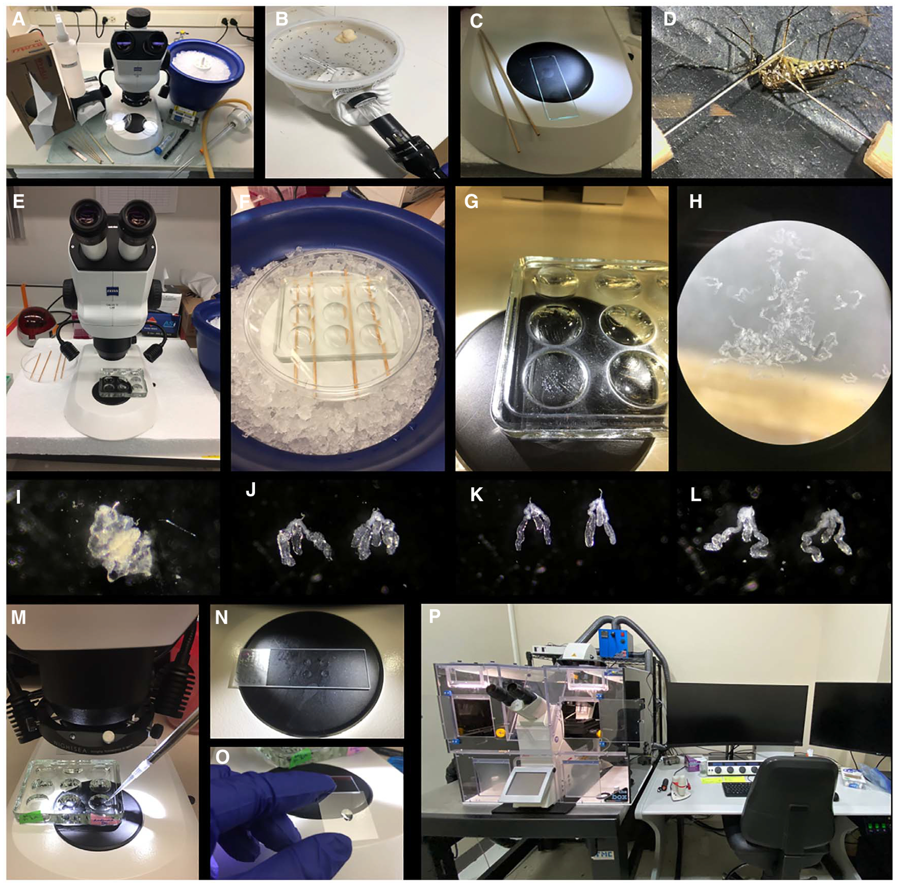
FIGURE 1.

Immunohistochemistry of mosquito salivary glands. (A) Dissection station. (B) Mosquito aspiration. (C) Fine needles and concave well slide for dissections (Step 2/3). (D) Aedes aegypti female mosquito dissection (Step 3). (E) Stereomicroscope setup. (F) While dissecting, salivary glands are deposited into a glass concave well plate and maintained on ice (Step 6). (G,H) Concave well plate with dissected salivary glands. (I) Ae. aegypti salivary glands surrounded by fat body tissue (Step 5). (J–L) Clean Ae. aegypti, Anopheles gambiae, and Culex quinquefasciatus salivary glands, respectively. (M) Washing step monitored under stereoscope. (N) Salivary glands are transferred to a drop of PBS onto a slide (Step 17). (O) Prolong Gold mounting media is added to a coverslip (Step 19). (P) Leica SP8 confocal microscope (Step 22).
