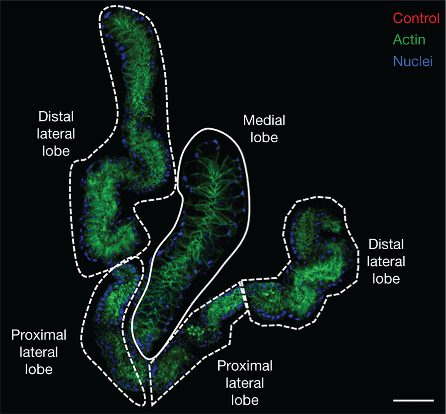
FIGURE 1.

Anatomy of salivary glands. Salivary glands of Aedes aegypti stained with mouse antibodies raised against adjuvant only. Distal and proximal lateral lobes are indicated with dashed white lines. The medial lobe is surrounded by a solid white line. Actin, stained with Phalloidin 488, and nuclei, stained with DAPI, are shown in green and blue, respectively. Scale bar, 50 μm.
