Figure 3.
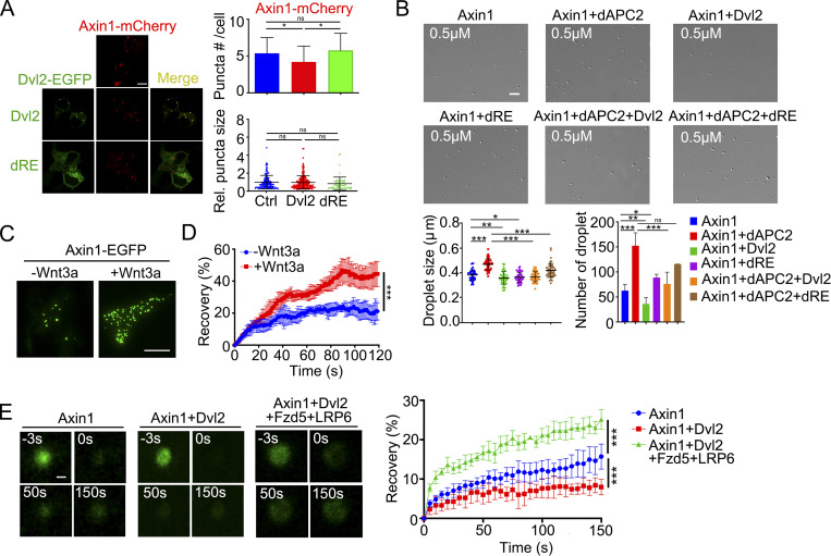
Dvl2 modulates the biophysical properties of Axin1 condensates. (A) Confocal images of Axin1-mCherry puncta when EGFP-tagged Dvl2 or Dvl2(dRE) were coexpressed in Dvl1/2/3 KO cells. The puncta size was normalized to that in the control group. (B) 0.5 µM Axin1 droplet formation induced by 10% PEG8000 after incubation with 0.5 µM dAPC2 and 0.5 µM Dvl2 or dRE. Quantification of droplet size and number are shown below. (C) TIRF images showing the membrane-associated Axin1-EGFP puncta in Axin1 KO cells with or without Wnt3a stimulation. (D) FRAP analysis of the membrane-associated Axin1-EGFP puncta detected by TIRF. (E) FRAP analysis of Alexa Fluor 488 NHS ester labeled Axin1 droplets incubated with or without the indicated components. The recovery of the fluorescent signal in Axin1 droplets was shown in the right. Statistical analyses were performed with the two-tailed unpaired t test. Data are shown as mean ± SD (n = 3). *, P < 0.05, **, P < 0.01, ***, P < 0.001. Scale bars in A and C, 10 µm; B, 2 µm; E, 0.2 µm.

