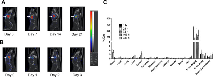
Figure 1.
Sagittal SPECT/CT images of an NRG mouse with a U251-Luc human GBM tumor in the right cerebral hemisphere of the brain (arrows) after CED of 1.0 MBq of (A) 177Lu-AuNPs or (B) 177Lu-MCPs (not conjugated to AuNPs). Images were obtained 0, 7, 14, and 21 d post-infusion for 177Lu-AuNPs and 0, 1, 2, and 3 d post-infusion for 177Lu-MCPs. (C) Percent injected dose/g (% ID/g) of 177Lu in the tumor-bearing right cerebral hemisphere, contralateral left hemisphere, or cerebellum and in the blood and other organs at selected times after CED of 1–2 MBq of 177Lu-AuNPs (4 × 10¹¹ AuNPs) in NRG mice. The y-axis was split to more clearly show uptake in organs with low % ID/g.

