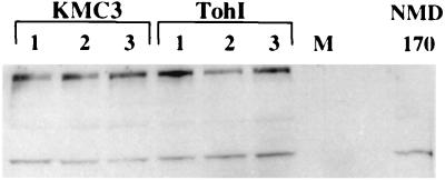
FIG. 3.
Western blot analysis of FHA expression. Whole-cell lysates from three cultures each of KMC3 and Tohama I (TohI), as well as a whole-cell lysate from NMD170 (an FHA− strain of B. pertussis), were analyzed by Western immunoblotting. FHA was detected with an FHA-specific goat polyclonal antibody. The upper high-molecular-weight band, missing in NMD170, represents FHA, while the predominant lower band is presumably a cross-reacting protein. M, rainbow marker (Amersham).

