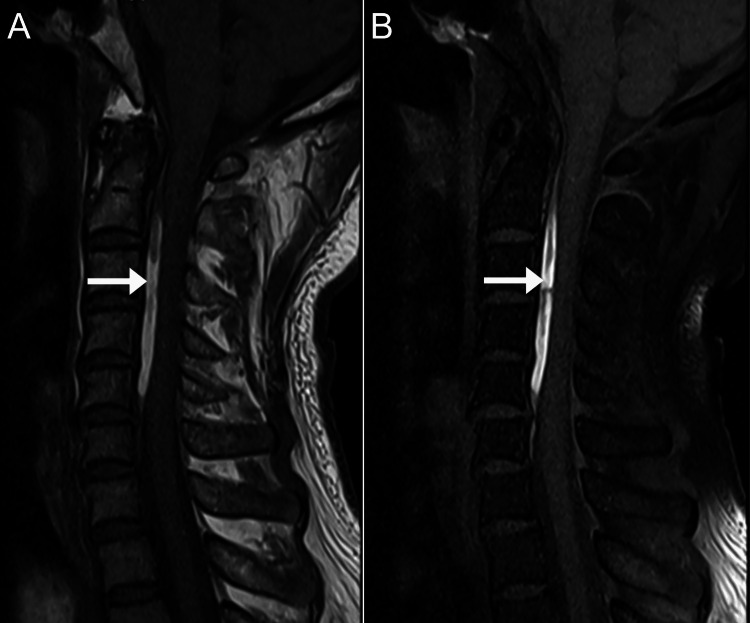
Figure 1. Magnetic resonance imaging of the cervical spine, mid-sagittal T1-weighted images.
A: Shows a linear hyperintense layer in the anterior epidural space spanning the vertebral levels C2 to C5 measuring 1.2 x 0.4 x 5.4 cm (arrow).
B: On a fat-suppressed sequence, this layer remains hyperintense (arrow), which suggests there is no macroscopic epidural fat within this layer.

