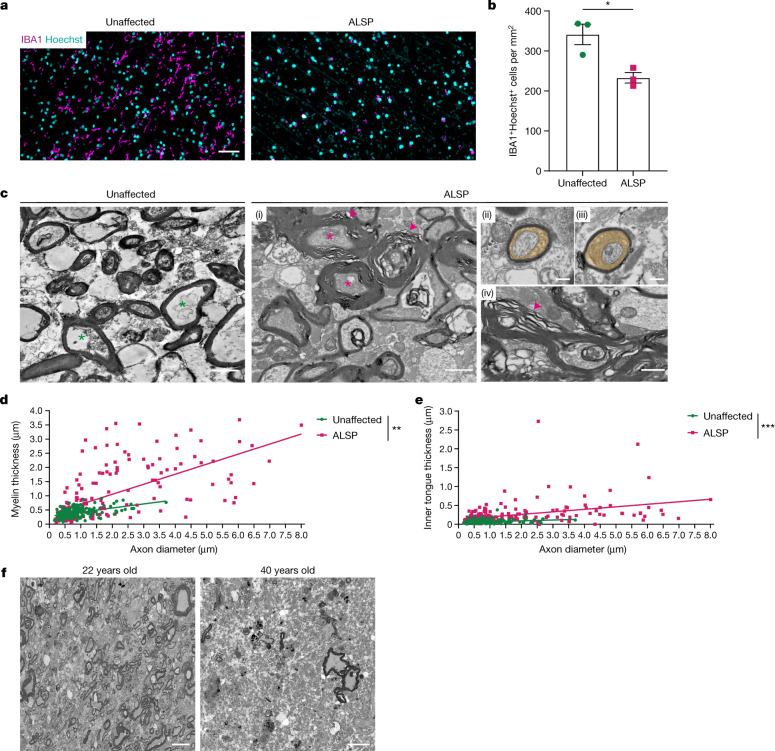
Fig. 4. Reduction of microglia in human white matter is associated with hypermyelination and demyelination.
a, Images of IBA1+ macrophages (magenta) in human frontal white matter from individuals with ALSP and unaffected age-matched individuals, counterstained with Hoechst (cyan). b, Mean IBA1+ cells per mm2 ± s.e.m. in samples from unaffected individuals and individuals with ALSP. n = 3 samples per group. *P = 0.0197, two-tailed unpaired Student’s t-test. c, Images of frontal white matter in unaffected and ALSP samples (i). Asterisks indicate axons of similar size, arrowheads indicate myelin abnormalities. Panels (ii) and (iii) show enlarged inner tongues in ALSP (orange). Panel (iv) shows myelin outfoldings and unravelling in ALSP (arrow). d, Myelin thickness versus axon diameter. n = 100 axons per sample, n = 2 samples per group. **P = 0.002, simple linear regression of slopes. e, Inner tongue thickness (µm) versus axon diameter. n = 100 axons per sample, n = 2 samples per group. ***P < 0.0001, simple linear regression of intercepts. f, Images of extent of demyelination of frontal white matter in individuals with ALSP; 22-year-old individual and 40-year-old individual. Scale bars, 0.5 µm (c (ii)–(iv)), 1 µm (c (i)). 10 µm (f) or 50 µm (a).

