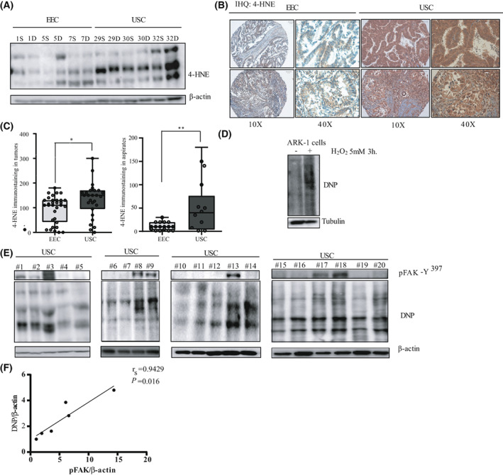
Fig. 4.

Enhanced oxidative stress markers in USC. (A) Representative western blot of oxidative stress marker 4‐HNE in 12 Endometrial Cancer samples (6 EEC and 6 USC). (n = 3 independent experiments). Β‐actin expression is used as a loading control. (B) Representative pictures of 4‐HNE immunostaining in two EEC samples and two USC samples. Pictures were taken at 10× and 40× magnifications. Scale bar: 100 μm. (C) 4‐HNE staining intensity score (immunohistochemistry) was calculated by a histoscore method (being 300 points the maximum immunoreactivity). Values are median ± CI (n = 3 independent experiments), statistical analysis was performed to compare 4‐HNE staining intensity in EEC and USC tumor samples and tumor aspirates. Statistical analysis of the obtained results shows a significant increase in 4‐HNE expression in USC tumors compared to EEC ones (Mann–Whitney U‐test; *P = 0.03) and in USC tumor aspirates compared to their EEC counterparts (Mann–Whitney U‐test; **P = 0.01). (D) Protein carbonylation was analyzed by western blot with anti‐DNP antibody in ARK‐1 cells that were either left untreated or stimulated with 5 mm of H2O2 for 3 h (n = 3 independent experiments). (E) Western blot with anti‐DNP in 20 USC samples (that were previously used for the kinome analysis; n = 3 independent experiments). (F) Spearman's rank correlation coefficient analysis comparing p‐FAK‐Y397/β‐actin vs. DNP/β‐actin protein levels in the six samples (numbers 15–20) of the western blot (Spearman r s = 0.94; P = 0.016).
