. 2022 Oct 3;22(1):102–111. doi: 10.1158/1535-7163.MCT-22-0375

Figure 2.

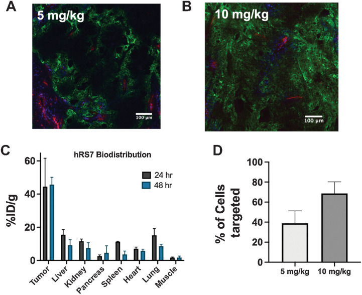
Figure 2. Organ, tissue, and cellular biodistribution. Tissue penetration of hRS7 is dose-dependent, with a 5-mg/kg dose showing heterogeneous distribution after 24 hours (A), while the clinical dose of 10 mg/kg penetrates deeper into the tumor (B). Blood vessels are imaged with anti-CD31 stain (red), intravenous Hoechst 33342 is shown in blue, and hRS7-AlexaFluor680 is in green. The high expression and rapid internalization results in efficient tumor uptake (n = 3; C). Flow cytometry of single-cell suspensions at 24 hours from three different tumors at each dose confirms a greater proportion of cells are hRS7-AlexaFluor680 positive at the higher dose (D). Data are shown as the mean and SD.

Organ, tissue, and cellular biodistribution. Tissue penetration of hRS7 is dose-dependent, with a 5-mg/kg dose showing heterogeneous distribution after 24 hours (A), while the clinical dose of 10 mg/kg penetrates deeper into the tumor (B). Blood vessels are imaged with anti-CD31 stain (red), intravenous Hoechst 33342 is shown in blue, and hRS7-AlexaFluor680 is in green. The high expression and rapid internalization results in efficient tumor uptake (n = 3; C). Flow cytometry of single-cell suspensions at 24 hours from three different tumors at each dose confirms a greater proportion of cells are hRS7-AlexaFluor680 positive at the higher dose (D). Data are shown as the mean and SD.