. 2022 Oct 3;22(1):102–111. doi: 10.1158/1535-7163.MCT-22-0375

Figure 4.

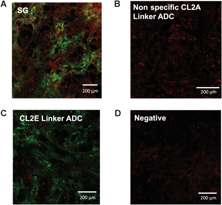
Figure 4. In vivo imaging of ADC distribution and payload-mediated DNA damage. Forty-eight hours following a 10-mg/kg dose of SG (A), nonspecific CL2A-SN38 ADC (B), hRS7-CL2E-SN38 (C), or uninjected mice bearing NCI-N87 xenografts, tissue was excised and imaged using an anti-Fc stain (green) or γH2AX DNA damage marker (red). SG shows the highest signal including cells lacking ADC targeting (bystander effects). Signal from the other ADCs was lower but above background.

In vivo imaging of ADC distribution and payload-mediated DNA damage. Forty-eight hours following a 10-mg/kg dose of SG (A), nonspecific CL2A-SN38 ADC (B), hRS7-CL2E-SN38 (C), or uninjected mice bearing NCI-N87 xenografts, tissue was excised and imaged using an anti-Fc stain (green) or γH2AX DNA damage marker (red). SG shows the highest signal including cells lacking ADC targeting (bystander effects). Signal from the other ADCs was lower but above background.