. 2022 Oct 3;22(1):102–111. doi: 10.1158/1535-7163.MCT-22-0375

Figure 5.

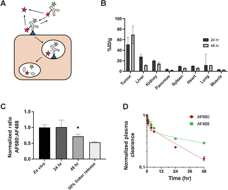
Figure 5. Dual-labeled ADC for quantifying extracellular release. By labeling hRS7 antibody with two residualizing dyes, AF488 (green star) attached directly to antibody via a stable amide linkage and SN38-AF680 (red star) connected via the CL2A hydrolysable linker, the location of payload release could be quantified. Extracellular released SN38-AF680 dye (red star) is unable to enter cells and washes out of the tumor, while intracellularly released payload is trapped (A). Biodistribution (% injected dose/gram) of the dual-labeled ADC (as measured by SN38-AF680 signal) shows high tumor uptake, consistent with intracellular release and residualization of the dye (B). The ratio of AF680 to AF488 indicates little extracellular release of the payload in NCI-N87 tumors. There is a significant drop in the AF680 to AF488 ratio between the ex vivo control and the ratio at 48 hours (P < 0.05; C). The decreased ratio at 48 hours is less than the positive control cells labeled with ADC following 50% SN38-AF680 release. This lower ratio can be attributed to the loss of SN38-AF680 in systemic circulation, where AF680 signal decreases faster than AF488 in the plasma due to deconjugation (D). Data are shown as the mean and SD for n = 3 mice at each timepoint. An outlying point at 24 hours in the lung (15.9, 16.5, and 73%ID/g), likely from blood clotting during processing, resulted in a large standard deviation at 24 hours.

Dual-labeled ADC for quantifying extracellular release. By labeling hRS7 antibody with two residualizing dyes, AF488 (green star) attached directly to antibody via a stable amide linkage and SN38-AF680 (red star) connected via the CL2A hydrolysable linker, the location of payload release could be quantified. Extracellular released SN38-AF680 dye (red star) is unable to enter cells and washes out of the tumor, while intracellularly released payload is trapped (A). Biodistribution (% injected dose/gram) of the dual-labeled ADC (as measured by SN38-AF680 signal) shows high tumor uptake, consistent with intracellular release and residualization of the dye (B). The ratio of AF680 to AF488 indicates little extracellular release of the payload in NCI-N87 tumors. There is a significant drop in the AF680 to AF488 ratio between the ex vivo control and the ratio at 48 hours (P < 0.05; C). The decreased ratio at 48 hours is less than the positive control cells labeled with ADC following 50% SN38-AF680 release. This lower ratio can be attributed to the loss of SN38-AF680 in systemic circulation, where AF680 signal decreases faster than AF488 in the plasma due to deconjugation (D). Data are shown as the mean and SD for n = 3 mice at each timepoint. An outlying point at 24 hours in the lung (15.9, 16.5, and 73%ID/g), likely from blood clotting during processing, resulted in a large standard deviation at 24 hours.