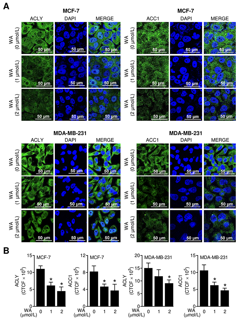
Figure 1.

WA treatment decreased protein levels of ACLY and ACC1 in human breast cancer cells. (A) Representative confocal images (60× oil objective magnification, scale bar = 50 μm) for ACLY and ACC1 proteins (green fluorescence) in MCF-7 or MDA-MB-231 cells following 24-hour treatment with DMSO (control) or WA. Nucleus was stained with DAPI (blue fluorescence). (B) Quantitation of corrected total cell fluorescence (CTCF) using ImageJ software. The results shown are mean ± SD (n = 3). Statistically significant (*P < 0.05) compared with the DMSO-treated control by one-way ANOVA followed by Dunnett’s test. The experiment was repeated two times and the results were consistent.
