Figure 5.

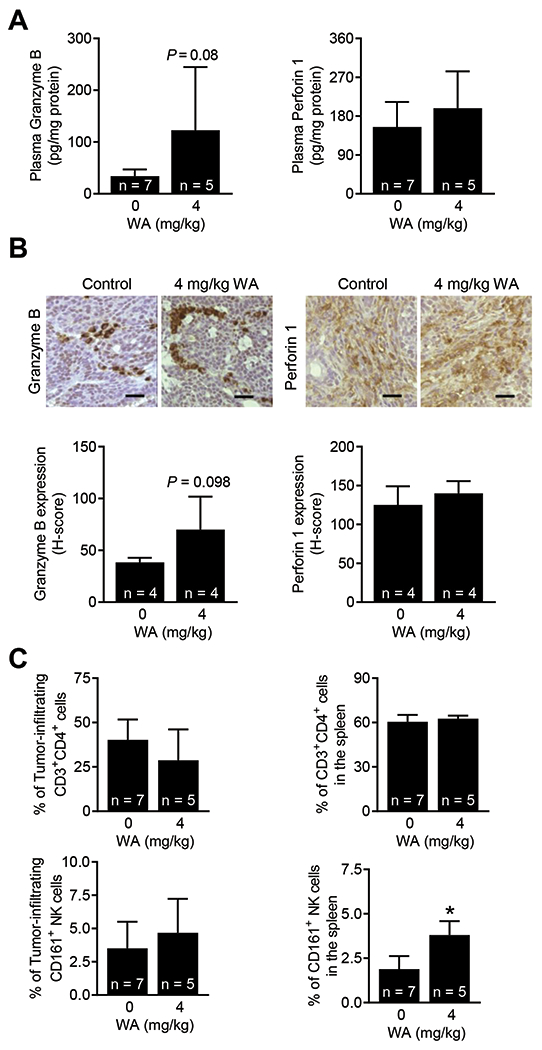
Effect of WA administration on immune cells/markers. (A) Quantification of Granzyme B and perforin 1 in the plasma of control and WA-treated rats. The results shown are mean ± SD (n = 5-7). Statistical significance (*P < 0.05) was determined by Student’s t-test. (B) Representative immunohistochemical images (200× magnification; scale bar = 50 μm) showing the expression of granzyme B and perforin 1 proteins in mammary tumors from a rat administered vehicle or WA. Quantification of tumor-infiltrating Granzyme B and perforin 1 are shown in lower panels. The results shown are mean ± SD (n = 4). Statistical significance (*P < 0.05) by Student’s t-test. (C) Quantification of immune cells in mammary tumors (left) and spleen (right). The results shown are mean ± SD (n = 5-7). Statistical analysis was done by Student’s t-test (*P < 0.05).
