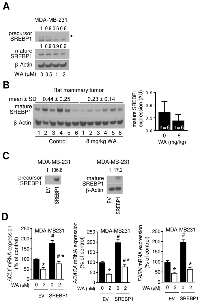
Figure 6.

WA administration inhibited SREBP1 expression. (A) Immunoblotting for precursor and mature forms of SREBP1 using lysates from MDA-MB-231 cells treated for 24 hours with DMSO (control) or the indicated doses of WA. The numbers above bands represent fold change of each protein relative to DMSO-treated control. (B) Immunoblotting for mature form of SREBP1 protein using mammary tumor lysates from control and WA-treated rats. The results shown are mean ± SD (n = 6). Statistical significance was determined by Student’s t-test. AU, arbitrary unit. (C) Immunoblotting for precursor and mature forms of SREBP1 using lysates from empty vector (EV) transfected or SREBP1 overexpressing MDA-MB-231 cells. (D) Real-time RT-PCR for ACLY, ACACA and FASN mRNA expression in EV and SREBP1 transfected MDA-MB-231 cells after 24 hours treatment with DMSO or 2 μmol/L WA. Results shown are mean ± SD (n = 3). Significantly different (P < 0.05) compared with DMSO-treated control (*) or between EV and SREBP1 cells at the same dose of WA (#) by one-way ANOVA followed by Bonferroni’s multiple comparisons test. The results were reproducible from replicate experiments.
