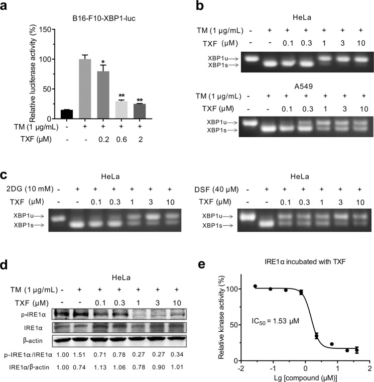
Fig. 2. TXF inhibits IRE1α kinase and RNase.
a B16-F10-XBP1-luc cells were treated with the indicated concentrations of TXF in the presence of TM (1 μg/mL). After 8 h, the cells were lysed and subjected to luciferase assay. *P < 0.05; **P < 0.01, versus vehicle group (n = 3), unpaired two-tailed Student’s t-test. b HeLa or A549 cells were treated with the indicated concentrations of TXF and TM (1 μg/mL), and c HeLa cells were treated with the indicated concentrations of TXF and 2DG or DSF for 8 h. XBP1 mRNA splicing was evaluated by semiquantitative RT-PCR. d HeLa cells were treated with the indicated concentrations of TXF and TM (1 μg/mL) for 8 h. The phosphorylation of IRE1α was evaluated by Western blotting. The band intensities were quantified by ImageJ software and the normalized ratio of p-IRE1α/IRE1α and IRE1α/β-actin were calculated. e Kinase activities measured after indicated concentrations of TXF were added to IRE1α for 1 h.

