Figure 3.

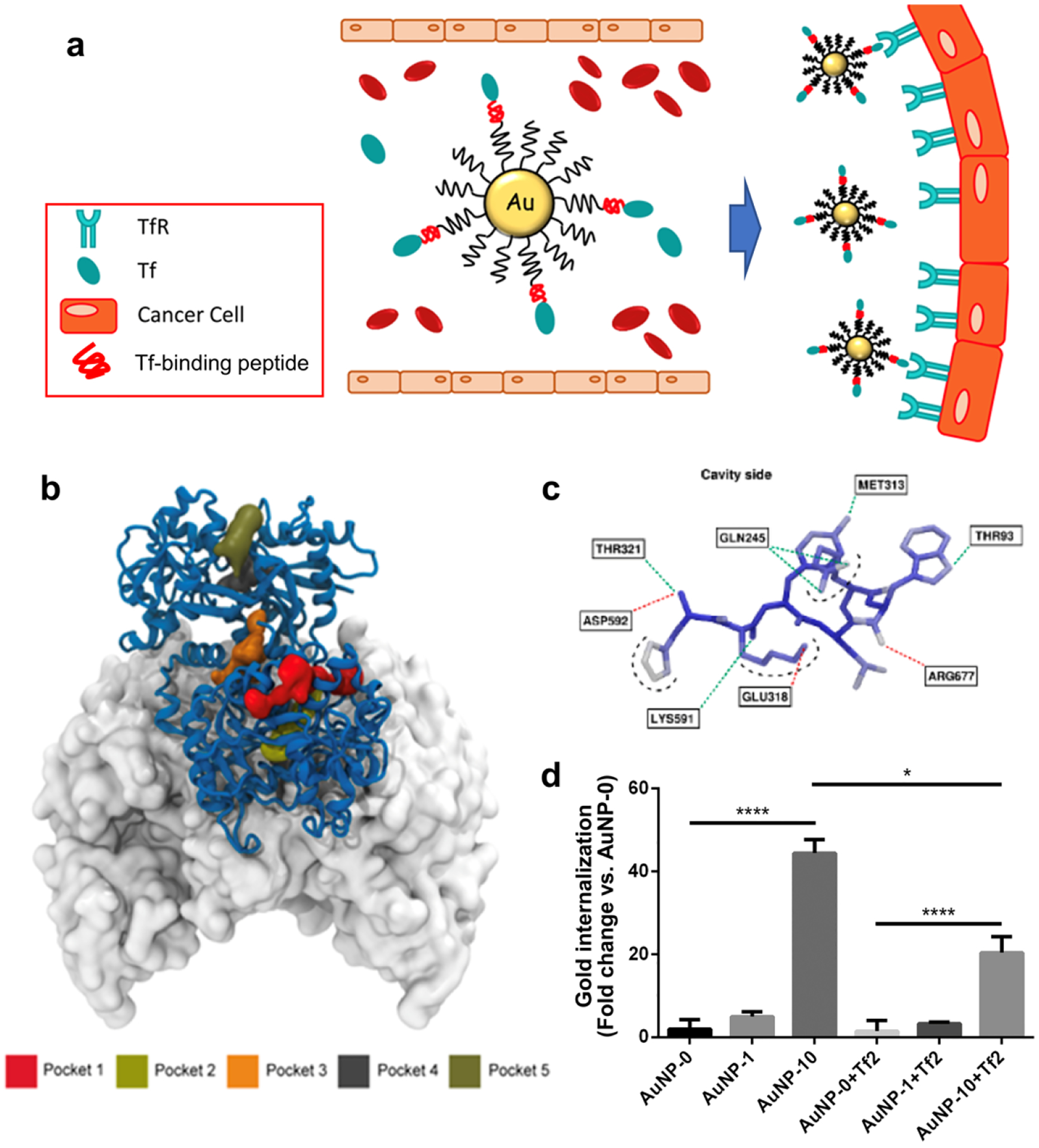
(a) Schematics Illustration of gold NPs targeting TfR overexpressed on cancer cells. (b) Five potential binding sites, Pocket 1 through 5, identified on the human Tf predicted by Fpocket. Tf is represented with the blue-ribbon diagram, and part of the ectodomain of the transferrin receptor dimer23 is rendered by its accessible surface. Due to the adequate volume for peptide and the distance from transferrin or iron binding site, Pocket 3 (orange) is chosen to dock the peptide. (c) 3D docked pose of the synthesized Tf-binding peptide (Tf2) created by coarse-grained molecular dynamics simulation. Atoms are colored according to their root mean squared displacement. Blue: rigid regions; red: flexible regions; green dashed lines: hydrogen bonds; red dashed lines: salt bridges; black dashed lines: solvent exposed atoms. (d) Cell uptake of gold NPs conjugated with Tf2. Various gold NPs were prepared using different percentage of PepN-Tf2 (0, 1, and 10% w/w). NPs were incubated in human plasma for 1 h at 37 °C and then with Mia PaCa-2 cells for 1 h at 37 °C in DMEM with (right, suffix +Tf2) or without (left) a Tf2. The results were normalized to the amount of internalized gold in AuNP-0. Reprinted from [201] with permission.
