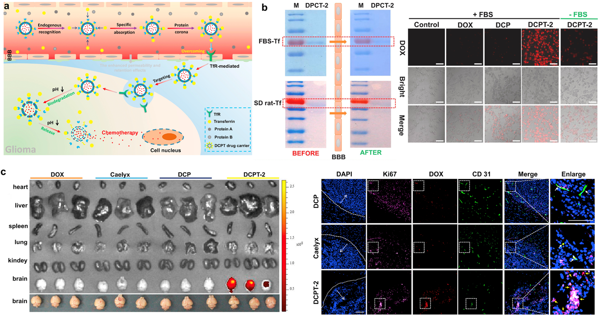
Figure 4.

Tf targeting in glioma by DOX-loaded, T10-coated COF NPs (DCPT) NPs. (a) Schematic illustration of endogenous Tf corona-mediated DCPT delivery across the BBB. (b) SDS-PAGE analysis of PC on DCPT-2 before and after passage through the in vitro BBB model (left). FBS-Tf: Formation of Tf corona on the surface of DCPT-2 mediated by Tf from the FBS. SD rat-Tf: Formation of Tf corona on the surface of DCPT-2 mediated by Tf from the SD rat serum. Cellular uptake of DOX and COF formulations incubated with U87 cells under different conditions (right). (c) Ex vivo imaging of DOX in main organs of glioma-bearing mice after intravenous injection of DOX, Caelyx, DCP (DOX-loaded COF, no T10) and DCPT-2 at 12 h (left). Immunofluorescence images of brain sections from orthotopic glioma mice after 12 h post-injection of DCP, Caelyx and DCPT-2, respectively. Blue: nuclei; purple: U87 cells; red: DOX; green: anti-CD31 labeled blood vessels. White arrows: co-localization of DOX and blood vessels; Yellow arrows: co-localization of DOX and glioma cells. Bar: 200 μm. Reprinted from [205] with permission.
