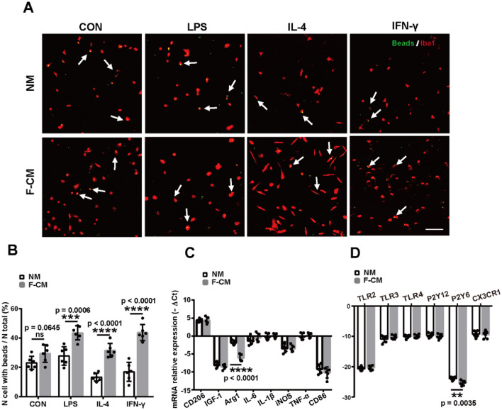
Fig. 4.
Phagocytosis and baseline levels of mRNA expression of primary cultured microglia isolated from the two groups. a Representative images of microglial (red) phagocytosed beads (green) after treatment with 100 ng/ml of LPS, 20 ng/ml IL-4 or 20 ng/ml of IFN-γ for 6 h. Scale bar = 100 μm. b The quantification of phagocytic ability. n = 6. c Baseline levels of gene expression. n = 5. d Baseline levels of microglial receptor mRNA expression. n = 6. **p < 0.01; ***p < 0.001; ****p < 0.0001; ns: no significant difference

