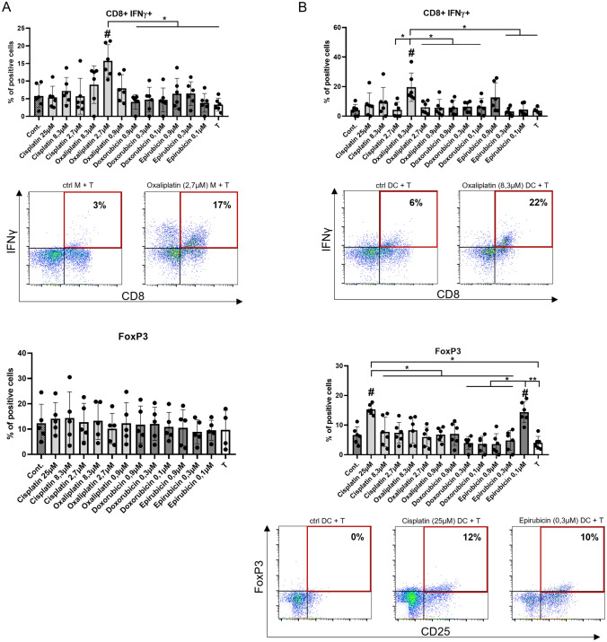
Fig. 5.
Chemotherapeutic agent-driven differentiation differently alter the T-cell-polarizing capacity of macrophages and dendritic cells. CD14+ monocytes were cultured with 50 ng/ml recombinant M-CSF (for macrophages) or 100 ng/ml IL-4 and 80 ng/ml GM-CSF (for dendritic cells) in the presence of cisplatin/oxaliplatin/doxorubicin or epirubicin for five days. On day five macrophages or dendritic cells were co-cultured with allogenous peripheral blood lymphocytes (PBL) at a monocyte-derived cell: T-cell ratio of 1: 10 at 37 °C.”T” sample indicates control consisting of only T cells without monocyte-derived cells. After three (CD8+ Tc) or nine days (CD4+ Treg) the T cells were stimulated with 1 μg/ml ionomycin and 20 ng/ml phorbol-myristic acetate (PMA) for 4 h, and the vesicular transport was inhibited. The ratio IFNγ producing cytotoxic T cells and CD4+ regulatory T lymphocytes (CD25+ IL-10+) were detected after the co-culturing of PBL with control or chemotherapeutics-conditioned macrophages (A) or dendritic cells (B). Representative dot plots of samples showing significant differences are shown. The cells' ratio for the measured molecules was calculated from at least five independent experiments + SD and In the statistical analysis, ANOVA followed by Bonferroni’s post hoc test was used for the comparison. The results were expressed as mean ± standard deviation. Differences were considered to be statistically significant at p < 0.05. Significance was indicated as #p < 0.05 compared to untreated control cells and as *p < 0.05 compared to the chemotherapeutic agent-treated counterparts

