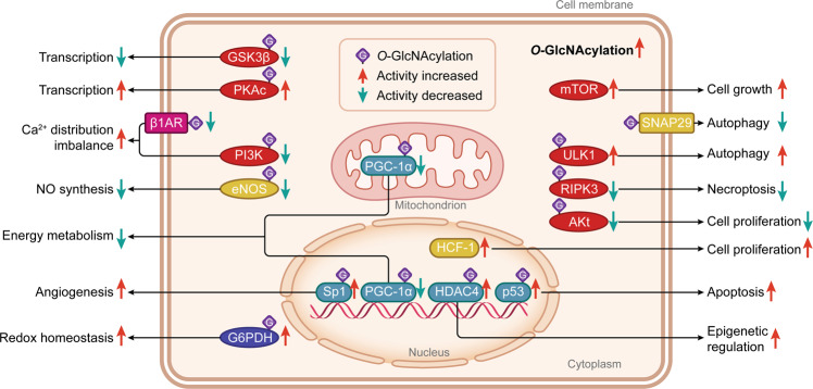
Fig. 1. The types, cellular distribution, and functions of proteins that could be O-GlcNAcylated in cells.
O-GlcNAcylated proteins, mainly located in the cytoplasm, nucleus, mitochondrion, and cell membrane, include the protein kinases (in red color), transcription factors and coactivators (in green color), metabolic proteins (in blue color), membrane receptors (in pink color), and other signaling molecules (in yellow color). They are responsible for essential activities with regard to epigenetic regulation, gene transcription, energy metabolism, cell cycle behavior, vascular function, Ca2+ distribution, NO production and redox homeostasis. Most of these proteins can be O-GlcNAcylated (in purple squares with the letter ‘G’) and their activities and functions can be changed directly. Differently, mTOR and SNAP29 are directly triggered by the higher intracellular O-GlcNAcylation levels. Proteins with enhanced activity after O-GlcNAcylation modification are indicated by red arrows, and proteins with decreased activity after O-GlcNAcylation modification are shown with green arrows. GSK-3β Glycogen synthase kinase-3β, PKAc PKA catalytic subunit, β1AR β1-adrenoceptor, PI3K Phosphoinositide 3-kinase, eNOS Endothelial nitric oxide synthase, G6PDH Glucose 6-phosphatedehydrogenase, PGC-1α Peroxisome proliferator-activated receptor-γ coactivator-1α, Sp1 Specificity protein 1, HDAC4 histone deacetylase 4, mTOR Mechanistic/mammalian target of rapamycin, SNAP29 Synaptosomal-associated protein 29, ULK1 Unc-51-like autophagy activating kinase 1, RIPK3 Receptor-interacting protein kinase 3, HCF-1 Host cell factor-1.

