
Figure 1.
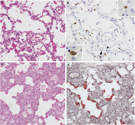
Representative histopathological findings in lung sections following hematoxylin–eosin (HE) (a, c, e) or Elastica–Masson Goldner staining (d). Immunohistochemistry was conducted to detect type A influenza virus nucleoprotein antigen (InfA-NP) (b). In situ hybridization AT-tailing (ISH-AT) (f–h) was also used. (a) Exudative phase of diffuse alveolar damage in Case 1. (b) InfA-NP antigen (brown) was detected in the nucleus (arrow head, upper inset) or cytoplasm (arrow, lower inset) of alveolar epithelial cells for Case 1. (c) Exudative phase of diffuse alveolar damage in case 3. (d) Elastica–Masson Goldner staining showed hyaline membrane formation (red) for case 3. (e) Proliferative phase of diffuse alveolar damage. The numbers of fibroblasts and myofibroblasts were increased in the alveolar septa and alveolar space in case 4. (f) ISH-AT using an antisense H5N1 nucleoprotein (NP) probe was able to detect H5N1 mRNA. (g) ISH-AT with a sense H5N1 NP probe detected H5N1 genomic RNA. (h) ISH-AT using an antisense rabies virus nucleocapsid probe as a negative control. Original magnifications, × 10 (c, d), × 20 (a, b), × 40 (b, inset, f–h).