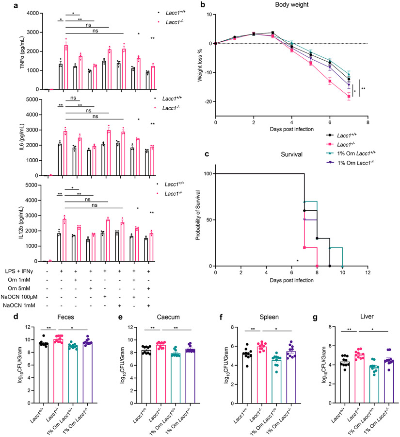
Fig. 2.
LACC1 inhibits proinflammatory cytokine signaling and protects from S. Typhimurium infection. a, ELISA measurements of TNFα, IL6 and IL12b secreted by BMDMs in the presence or absence of immunostimulant (LPS + IFNγ), exogenously supplied LACC1 product L-Orn (1, 5 mM), and/or the conjugate base of LACC1 product HNCO, NaOCN (100 μM, 1 mM). b, Body weight of each mouse was monitored daily after S. Typhimurium infection with or without 1% L-Orn administration, and the body weight loss was depicted as the percentage (%) compared to the initial body weight (n = 10). c, Mouse survival curves (n = 10). d, Colony-Forming Units (CFUs) in fecal pellets were measured at day 4 after infection. e, f, g, CFUs in caecum, spleen and liver, respectively, were measured at day 6 after infection. The mean and SEM (error bars) are derived from three (Fig. 2a) or ten (Fig. 2b-g) biological replicates (n = 3 or n = 10). Statistical significance (two-tailed t-test) compared to control (Ctrl): *P<0.05; **P<0.01; ns, not significant.

