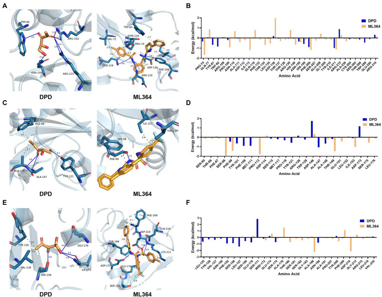
Figure 4.
The interactions and per-residue free energy decomposition between DPD or ML364 with DPD/AI-2 receptors. (A) The interactions between DPD or ML364 with LuxP protein of V. Campbellii. (B) The per-residue energy decomposition between LuxP with DPD or ML364. (C) The interactions between DPD or ML364 with PctA protein of Pseudomonas aeruginosa. (D) The per-residue energy decomposition between PctA with DPD or ML364. (E) The interactions between DPD or ML364 with TlpQ protein of P. aeruginosa. (F) The per-residue energy decomposition between TlpQ with DPD or ML364. Protein-ligand interactions are colored as following: blue solid line, hydrogen bond; dash line, hydrophobic interaction; cyan solid line, halogen bond.

