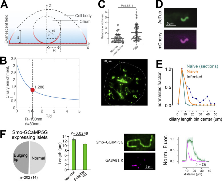
Figure S2.
Characterization of a cilia-targeted Ca2+ sensor. (A and B) Principle for determining the enrichment of Smo-GCaMP5G-mCh within the primary cilium of islet cells imaged by TIRF microscopy. With a radius of the cilium of 100 nm and an evanescent wave penetration depth (d) of 80 nm, we estimate that a ratio of cilia-to–plasma membrane fluorescence >1.288 indicate cilia enrichment. (C) Quantifications of fluorescence intensities of Smo-GCaMP5G-mCh from the plasma membrane and cilia of islet cells. The average cilia-to–plasma membrane fluorescence ratio is 2, which indicate strong cilia enrichment (means ± SEM; n = 113 soma and 95 cilia; Student’s two-tailed unpaired t test). (D) Immunofluorescence staining of a MIN6 cells expressing Smo-GCaMP5G-mCherry using antibodies against acetylated tubulin (green) and mCherry (magenta). (E) Measurements of cilia length in islets cells from mouse pancreatic sections (green), isolated mouse islets (red), and mouse islets transduced with Smo-GCaMP5G-mCh for 48 h (black). (F) Quantification of cilia morphologies in islet cells expressing Smo-GCaMP5G-mCh (means ± SEM; n = 202 cilia from 14 islets; two-tailed unpaired Student’s t test). Images show an example of the distribution of GABAB1 receptors in a cilium expressing Smo-GCaMP5G-mCh and quantification of the distribution along the cilia of 23 islet cells.

