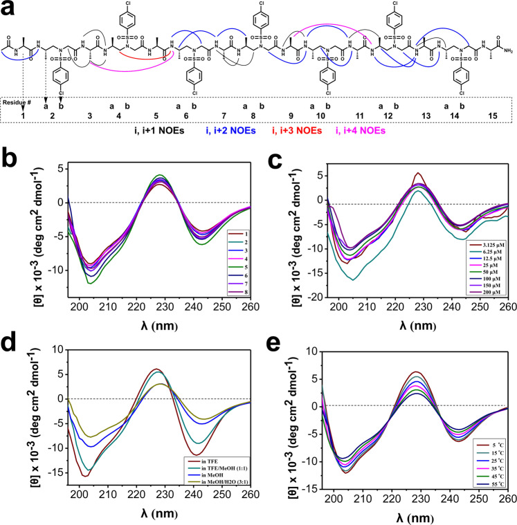
Fig. 4. Solution structures of foldamers.
a Summary of detected NOESY cross-peaks (200 ms mixing time) of foldamer 8 between protons on nonadjacent residues in CD3OH (4 mM concentration, 10 °C). Four types of NOEs are displayed in different color. Each l-sulfono-γ-AA peptide unit is considered as two residues since it is equal to two α-amino acids in length. b CD spectra of compounds 1‒8 (100 μM) measured at room temperature in CH3OH. c CD spectra of compound 8 in CH3OH at various concentrations at room temperature. d CD spectra of compound 8 (100 μM) in various solvents at room temperature. e CD spectra of compound 8 (100 μM) in CH3OH at various temperatures.

