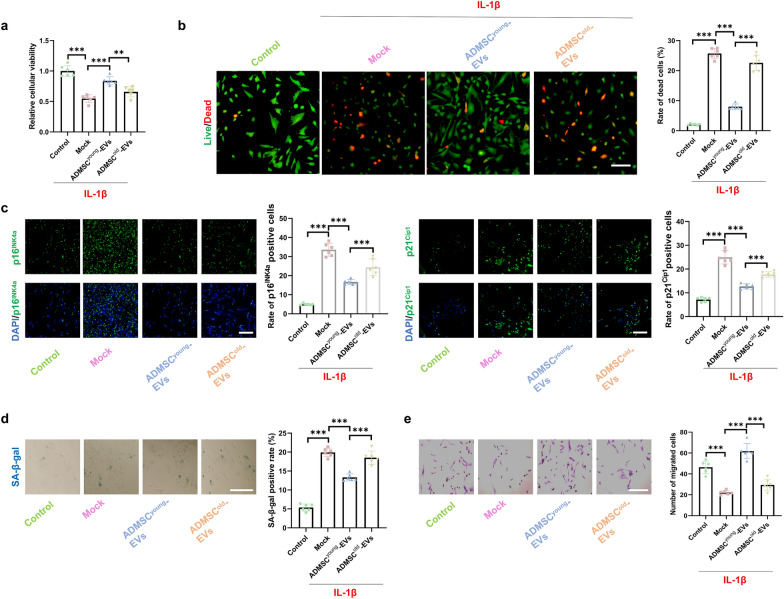
Fig. 5.
ADMSCyoung-EVs, but not ADMSCold-EVs, alleviated cell death and cellular senescence of tenocytes treated with IL-1β. a The cellular viability in the control, IL-1β, IL-1β + ADMSCyoung-EVs and IL-1β + ADMSCold-EVs tenocyte groups, n = 6. b Live/dead assay and rate of dead cells in the control, IL-1β, IL-1β + ADMSCyoung-EVs and IL-1β + ADMSCold-EVs tenocytes groups, n = 6. Live cells are marked green, and dead cells are marked red. Scale bar = 50 μm, n = 6. c Immunofluorescence staining of p16INK4A (green), p21CIP1 (green) and nuclear (DAPI, blue) in the control, IL-1β, IL-1β + ADMSCyoung-EVs and IL-1β + ADMSCold-EVs tenocytes groups. Scale bar = 200 μm, n = 6. d SA-β-gal activity and the percentages of SA-β-gal-positive cells in ADMSCyoung and ADMSCold. Scale bar = 50 μm, n = 6. e Images and quantification of migrated tenocytes in the control, IL-1β, IL-1β + ADMSCyoung-EV and IL-1β + ADMSCold-EV groups. Scale bar = 50 μm, n = 6. Data are presented as the mean ± SD (***: P < 0.001)

