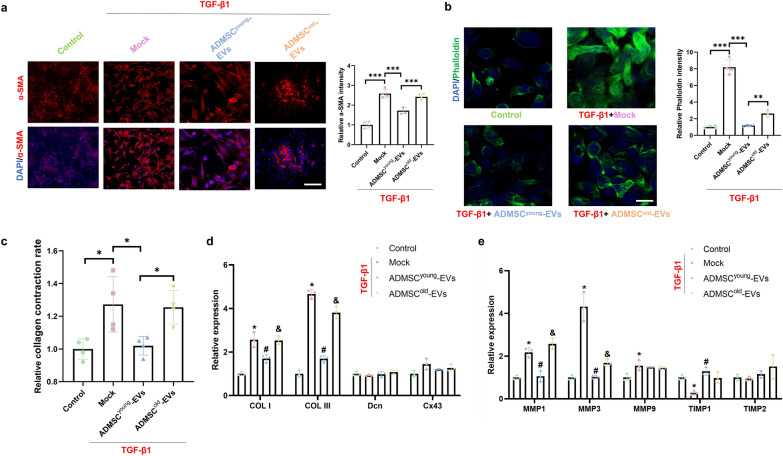
Fig. 6.
ADMSCyoung-EVs, but not ADMSCold-EVs, alleviated pathological ECM remodeling of tenocytes induced by TGF-β1. a Immunofluorescence staining and relative intensity of α-SMA (red) and nuclei (DAPI, blue) in the control, TGF-β1, TGF-β1 + ADMSCyoung-EV and TGF-β1 + ADMSCold-EV tenocyte groups. Scale bar = 200 μm, n = 4. b Immunofluorescence staining and relative intensity of phalloidin (green) and nuclei (DAPI, blue) in the control, TGF-β1, TGF-β1 + ADMSCyoung-EV and TGF-β1 + ADMSCold-EV tenocyte groups. Scale bar = 200 μm, n = 4. c Relative collagen contraction rate in the control, TGF-β1, TGF-β1 + ADMSCyoung-EV and TGF-β1 + ADMSCold-EV tenocyte groups, n = 4. d–e Relative expression of collagen formation- and degradation-related genes in the control, TGF-β1, TGF-β1 + ADMSCyoung-EV and TGF-β1 + ADMSCold-EV tenocyte groups, n = 3. Data are presented as the mean ± SD (*: control vs. TGF-β1 treatment; #: TGF-β1 treatment vs. TGF-β1 + ADMSCyoung-EVs treatment; &: TGF-β1 + ADMSCyoung-EVs vs. TGF-β1 + ADMSCold-EVs treatment groups, P < 0.05)

