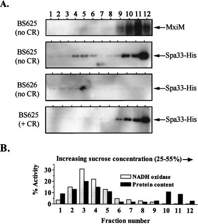
FIG. 5.
Spa33 distribution within the cellular envelope as determined by sucrose density gradient centrifugation. Strains were grown with arabinose prior to the isolation and fractionation of total cell envelope preparations. (A) Immunoblot analysis of samples isolated from strains BS625 (spa33-1/PBAD-spa33+-his) and BS626 (virulence plasmid-cured strain bearing the PBAD-spa33+-his fusion in trans) using anti-MxiM and anti-His antibodies. Equivalent aliquots of every fraction isolated from the Congo red (+ CR)-induced and -uninduced BS625 cultures were examined. For the BS626 (no CR) sample, five times more sample was analyzed per fraction. Exposure times for each Spa33-His blot are the same. The MxiM distribution patterns for strains BS625 (+ CR) and BS626 (no CR) were identical to that observed for BS625 (no CR) and for that reason are not shown. (B) NADH oxidase activity and total protein content in envelope fractions isolated from the BS625 (no CR) culture. The total percentage corresponds to the amount of NADH oxidase activity or protein per fraction divided by the total amount determined for each gradient. The NADH oxidase and protein distribution patterns for BS625 (+ CR) and BS626 (no CR) were identical and for that reason are not included.

