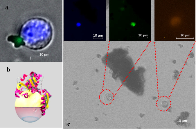
Fig. 4. Images of cancer cells captured by MFN from blood samples.
a HCT116 cell captured by MFN conjugated with Cy5. Cell visualized by nuclear staining with Hoechst reagent (blue) and Cy5 (green). b Schematic of the MFN conjugated with Tf/Ab. c Immunocytochemistry method based on Fluorescein Isothiocyanate-labeled anti-cytokeratin (green), and Hoechst (blue) nuclear staining was applied to identify and enumerate CTCs captured by MFNs. PE-labeled anti-CD45 (red) and Hoechst (blue) nuclear staining was used to identify leukocytes.

