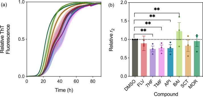
Fig. 3. The flavone derivatives affect the α-synuclein secondary nucleation step differentially.
a Normalised change in ThT fluorescence when 20 µM monomeric α-synuclein was incubated with 50 nM preformed seed fibrils at pH 4.8 and 37 °C in the absence (DMSO control (black)) and presence of 10 µM (0.5 molar equivalents, relative to monomeric protein) of flavone derivatives (flavone (red), 5,6,7-trimethoxyflavone (magenta), 7-hydroxyflavone (purple), apigenin (blue), baicalein (light green, which speeds up the process), scutellarein (brown), morin (dark green)). Yellow overlay indicates corresponding generalised logistic fit (Eq. 2). b Rate of α-synuclein fibril amplification, normalised relative to the DMSO control. Error bars represent the standard error of the mean of three experimental replicates each containing three technical replicates. Statistical analyses represent two-way ANOVA results where **P ≤ 0.0021.

