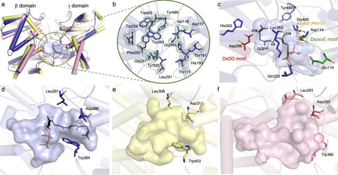
Fig. 2. Structure of Tpn2.
a Overall βγ didomain structure of Tpn2 (PDB ID 7XKX, blue) aligned with PtmT2 (PDB ID 5BP8, yellow) and Rv3377c (PDB ID 6VPT, pink). The active site pocket of Tpn2 is circled in green. b Hydrophobic residues found in the active site pocket of Tpn2. The σA-weighted difference (mFo − DFc) omit map for Tpn2 with a 3σ contour is shown in green mesh. c Proposed binding mode of GGPP in the active site of Tpn2. C14 of GGPP lies 3.2 Å away from the carboxylate side chain of D296. G485, and the corresponding D502 in PtmT2, lie 3.6 Å from C19 of GGPP. Comparisons of the active site pockets of Tpn2 (d), PtmT2 (e), and Rv3377c (f).

