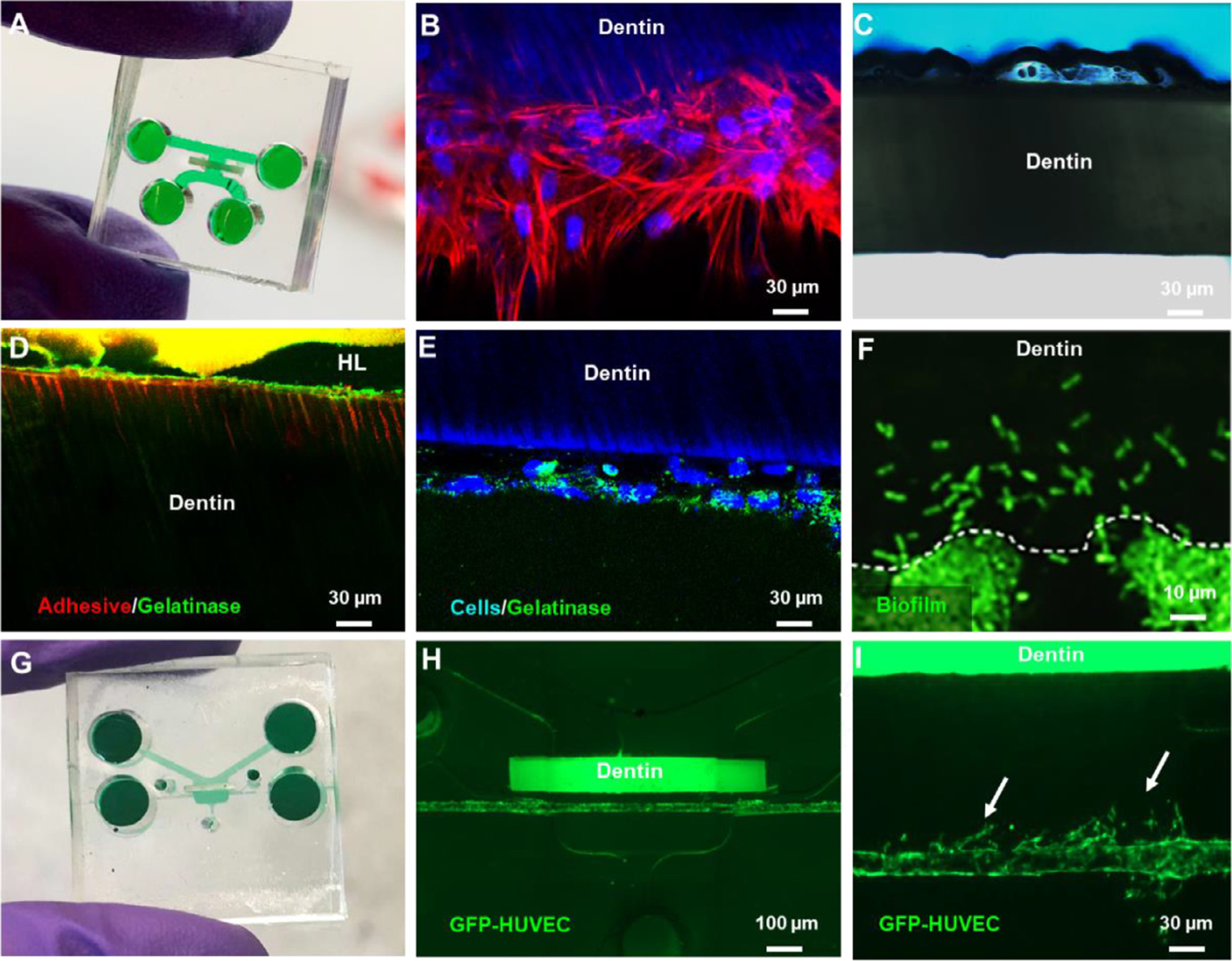
Fig. 5.

Applications of the Tooth on-a-chip to test dental materials. (A) Microfluidic device comprised of two parallel chambers separated by a fragment of native dentin. (B) Stem cell from apical papilla (SCAPs) seeded on-chip after 7 days protrude cytoplasmic processes into the dentin tubules akin to odontoblasts. (C) Interaction of phosphoric acid and native dentin recorded in real-time. (D) Tooth-on-a-chip devices were seeded with cells, and after 48 h, fluorescein-conjugated gelatin showed gelatinolytic activity in the hybrid layer and dentin tubules. (E) The gelatinolytic activity was co-localized with cell cytoplasm suggesting a role for cells in the enzymatic degradation of the hybrid layer. Reproduced with permission [77] (F). Streptococcus mutans seeded on-chip to evaluate real-time biofilm formation and interactions with pulp cells. (G) Tooth-on-a-chip for vasculature studies. (H) The device has a chamber on the ‘pulp side’ filled with collagen, seeded with mesenchymal stem cells and GFP-HUVECs. After 24 h, a pericyte-supported blood vessel is engineered with a controllable distance from the dentin. (I) Angiogenic sprouts toward the dentin on-chip.
