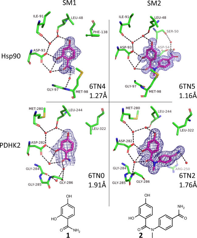
Fig. 1. Crystal structures of the starting materials bound to the two proteins.
Shown are the compounds (magenta carbon atoms) and water molecules and amino acids from the proteins that are within 3.5 Å of the compound (with green carbon atoms) and with red oxygen, blue nitrogen and yellow sulfur atoms. 2Fo–Fc electron density contoured at 1σ for the ligand is shown in dark blue (see Supplementary Fig. 6 for omit maps); hydrogen bonding between ligand, protein and solvent is shown with black dashed lines; pdb code and resolution shown for each structure.

