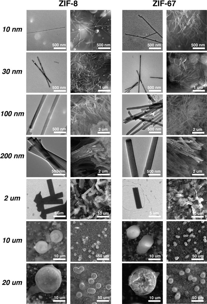
Fig. 4. Electron microscopy images of isolated nano- and micro-structures.
Isolated structures by dissolution (left columns) and on conducting tape supports (right columns) for both ZIF-8 and ZIF-67, respectively. For dissolved structures, images for sizes 10 nm to 2 μm are from transmission electron microscopy (TEM), while images for sizes 10 and 20 μm are from scanning electron microscopy (SEM). Structures on conducting tape supports are all analyzed by SEM.

