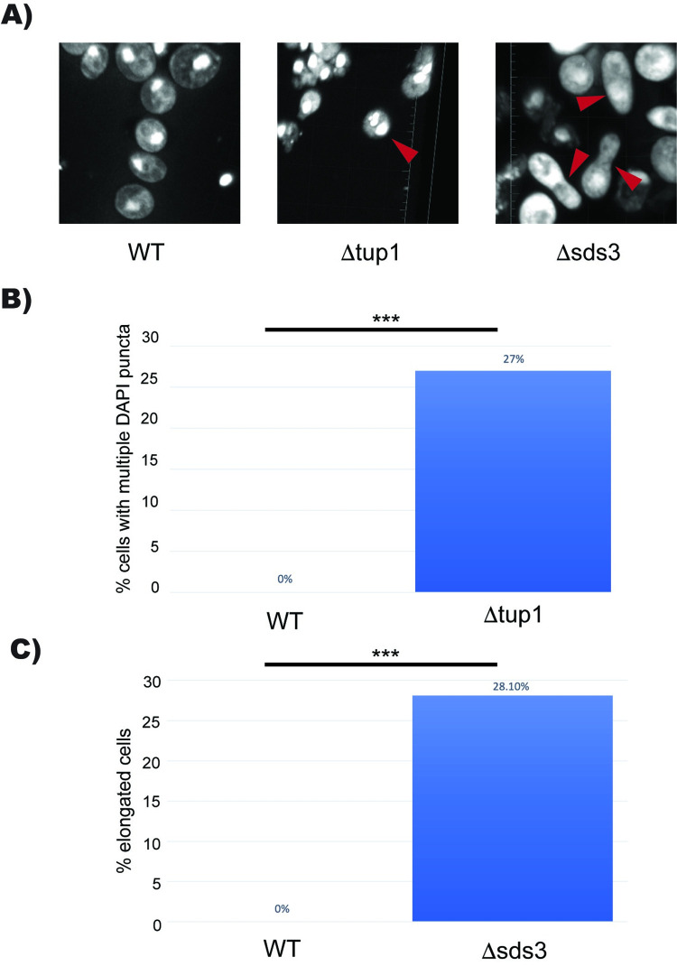
Fig 6. Deletion of Tup1 or Sds3 Leads to Morphology Defects in Stationary Phase.
(A) Fluorescence microscopy of DAPI stained nuclei across the genome at 67x magnification in wild type, Δtup1, and Δsds3 yeast. (B) Quantification of Δtup1 cells with multiple DAPI puncta. WT n = 100, Δtup1 n = 48. (C) Quantification of peanut-shaped cells in Δsds3 yeast. WT n = 100, Δsds3 n = 64. *** p < 0.05.

